

Staining criteria, collection and number of cells evaluated to identify micronuclei in buccal mucosa cells of agricultural workers exposed to pesticides
Criterios de tinción, recolección y número de células evaluadas para identificar micronúcleos en células de mucosa bucal de trabajadores agrícolas expuestos a pesticidas
Mexican Journal of Medical Research ICSA
Universidad Autónoma del Estado de Hidalgo, México
ISSN-e: 2007-5235
Periodicity: Semestral
vol. 9, no. 18, 34-38, 2021

Abstract: Although there are several publications that refer to the basic criteria established for the micronucleus (MN) test in exfoliated buccal cells, there is still a difference in the quantity, method of obtention and staining of the cells for the evaluation of the presence of micronuclei. Objective. Identify the criteria for evaluation of MN in oral mucosa cells exposed to pesticide in investigations carried out. Material and methods. A systematic review was carried out on the internet, based on articles published in Crossref, JCR, Scopus, PubMed, Google academic, using keywords such as: micronuclei, buccal mucosa cells, genotoxic damage, pesticides and biomarker. Results. In the six selected articles, four presented statistically significant values with the presence of MN when comparing the exposed groups with respect to the control groups, and the criteria for staining, collection and number of cells evaluated to identify micronuclei were very varied. Conclusions. It is important to follow validated and standardized protocols for the MN oral cytoma assay. Being considered in all the parameters suggested in the protocol will increase the reliability of the studies and will give the possibility of comparing the results obtained.
Keywords: Micronucleus, biomarker, genotoxic damage, buccal mucosa cells, pesticides.
Resumen: Aunque existen varias publicaciones que refieren los criterios básicos establecidos para la prueba de micronúcleos (MN) en células bucales exfoliadas aún hay diferencia en la cantidad, método de obtención y tinción de las células para la evaluación de presencia de micronúcleos. Objetivo. Identificar los criterios para evaluación de MN en células de mucosa oral de expuestos a pesticidas en investigaciones realizadas. Material y métodos. Se realizó una revisión sistemática en la red de internet, con base en artículos publicados en crossreff, JCR, Scopus, PubMed, Google académico, mediante palabras clave como: micronúcleos, células de mucosa bucal, daño genotóxico, pesticidas y biomarcador. Resultados. En los seis artículos seleccionados cuatro presentan valores estadísticamente significativos con presencia de MN al comparar los grupos expuestos respecto a los grupos control, y los criterios de tinción, recolección y número de células evaluadas para identificar micronúcleos son muy variados, Conclusiones. Es importante seguir los protocolos validados y estandarizados para el ensayo de MN citoma bucal. Al ser considerado en todos los parámetros sugeridos en el protocolo aumentará la fiabilidad de los estudios y dará la posibilidad de comparar los resultados obtenidos.
Palabras clave: Micronúcleos, biomarcador, daño genotóxico, células de mucosa bucal, pesticidas.
INTRODUCTION
In the first studies conducted in the 1980s, exfoliated buccal mucosa cells were used to assess the genotoxic effects of betel- quid nuts and chewing tobacco.1-6 Most studies showed higher frequencies of micronucleus (MN) at the site within the oral cavity where the quid or tobacco mixture was maintained compared to the opposite control site. The buccal cell MN assay was also used to study cancerous and precancerous lesions and to monitor the effects of a number of chemopreventive agents.7-9
It is notable that the first Stich and Rosin studies conducted between 1983 and 1984 had higher reference MN frequencies than subsequent studies. This may have been due to the lack of defined scoring criteria and a relatively small number of cells analyzed (in some cases less than 500). Since then, published biomonitoring studies using the MN assay on buccal mucosa cells have investigated the effects of multiple factors including environmental and occupational exposures, radiation therapy, chemoprevention, vitamin supplementation trials, lifestyle habits, cancer, and other diseases.10
An important aspect of the MN assay in buccal mucosa cells is the criteria for identifying and scoring cells. The five publications by Stich and Rosin1,2,8,11,12 refer to the basic criteria established for MN that were initially described by Heddle.13 However, criteria to identify cells for inclusion in the MN frequency count were not provided. Other authors refer to Heddle's criteria as such, or with minor modifications. The criteria developed by Tolbert et Alabama14,15 for choosing cells are the most used. They consist of the following parameters for cell inclusion in the cells to be scored: (a) intact cytoplasm and relatively flat cell position on the slide; (b) little or no overlap with adjacent cells; (c) little or no dirt; and (d) normal and intact nucleus,smooth and distinct nuclear perimeter.15
Some meta-nuclear changes indicative of cytotoxicity when analyzing the micronucleus assay in the cells of the oral mucosa, such as picnosis, carolysis and karyorrhexis. The approach is very important because cytotoxicity is a confounding factor for mutagenesis13,17 (a) rounded smooth perimeter suggesting a membrane; (b) less than a third of the diameter of the associated nucleus, but large enough to discern shape and color; (c) Feulgen positive, that is, rose in bright illumination field; (d) staining intensity similar to that of the nucleus; (e) core-like texture; (f) same focal plane as the nucleus; and (g) absence of overlap or bridge to the nucleus.15
Therefore, the objective of this review was to identify and determine the criteria to quantify MN in oral mucosa cells from previously conducted research.
MATERIAL AND METHODS
The review includes studies carried out on the publications of different investigations in humans and that specify the criteria for identifying micronuclei in cells of the oral mucosa. The literature search included the following keywords: "micronuclei" buccal mucosa cells, biomonitoring, biomarker, genotoxic damage, pesticides.
Searches were performed in literature databases including crossreff, JCR, Scopus, PubMed, Google academic in March 2020.
Inclusion and exclusion criteria
The inclusion criteria were biomonitoring human research using the buccal mucosa micronucleus test with limitation of having used Feulgen staining and specifically in exfoliated buccal mucosa cells, not including lymphocyte tests.
The extracted data included, research methodology, number and method of counting and identification, comparison between differences, number of participants and condition, during study follow-up.
The only exclusion criteria was the lack of clarity of the micronucleus identification criteria in the carried out studies.
RESULTS
Search for articles and data collection.
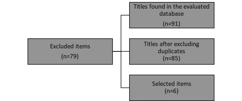
Our search retrieved 91 articles, of which 84 were articles made to a population that presented another type of exposure. In the end, six articles with results related to micronuclei in buccal mucosa cells of agricultural workers exposed to pesticides (specifying the criteria for staining and identification of micronuclei).
In the six selected articles, four presented statistically significant values with the presence of MN when comparing the exposed groups with respect to the controls, and the criteria for staining, collection and number of cells evaluated to identify micronuclei were very varied.
Characteristics of the studies.
Of the seven full articles evaluated for micronuclei in buccal mucosa cells of agricultural workers exposed to pesticides as a result, one was excluded from the systematic review because this article was a compilation of results from four other articles. Therefore, six studies were selected for qualitative analysis.
Of which two articles refer to having carried out the collection of samples through cytobrush, two with a wooden spatula and two with a toothbrush, also in the sample staining method it is different three used shiff reagent, one Giemsa and one of staining that is mentioned in these articles as a specific DNA staining called DAPI (dimethyl, diphenylidol, dichloride) and the latter with acridine orange. Regarding the total cell count, three counted 2,000 per person, 1-3,000 per person and 2 out of 1,000 per study subject. The standardized MN buccal assay protocol clearly states that 2,000 buccal cells stained with specific DNA staining must be scored for reliable results.15
DISCUSSION
There are several advantages to using the MN frequency in exfoliated buccal cells, such as the specificity to detect the effects of exposure to inhaled or ingested genotoxic agents, and the easy storage of samples, before and after processing, in fixative solution or as fixed slides at room temperature.16
Of the total number of articles reviewed there is a great variety with respect to validated and standardized protocols, others do not consider them, although they do mention the authors of this technique and only mention that they make some modifications, of the discarded articles which carry out the tests. Other types of population such as hairdressers, smokers, gasoline dispensers to name a few, there is still more variability in the collection methods, staining, and number of cells evaluated.
| Title | N° of Cells Evaluated | Collection Object | Staining method | |
| Cytotoxic and genotoxic effects of pesticide exposure in coffee workers in the Jarabacoa region, Dominican Republic. 17 | 2000 cells | Wet wooden spatula | Schiff's reagent (Sigma-Aldrich, Steinheim, Alemania) | |
| Cytogenetic biomonitoring of Brazilian workers exposed to pesticides: Analysis of micronuclei in buccal epithelial cells of soybean producers.18 | 1000 cells | Cytobrush | Giemsa's solution to 2% | |
| Micronuclei in buccal and peripheral blood lymphocytes epithelial cells from Polish farmers exposed to pesticides19 | 2000 cells | Toothbrush | 4, 6-di-amidino-2-fenilindol dihydrochloride (DAPI) | |
| Biomonitoring of agricultural workers exposed to pesticides mixtures in the state of Guerrero, Mexico, with kite testing and micronucleus test. 20 | 3000 cells | Wet wooden spatula | Schiff's reagent | |
| Results of the buccal micronucleus cytoma test in a group exposed and not exposed to pesticides. 21 | 1000 cells | Toothbrush | Schiff's reagent | |
| Biochemical and genotoxic effects in women exposed to pesticides in southern Ecuador.22 | 2000 cells | Cytobrush | Acridine Orange |
Pesticides are widely used worldwide, and pesticides exposure remains a major environmental health problem. Of significant importance are the potentially dangerous effects on occupationally exposed subjects. Although the genotoxic potential of pesticides is believed to be low, genotoxic monitoring in farm worker populations could be a useful tool in estimating the risk of long-term health effects such as cancer and adverse reproductive health outcomes.23,24
The systematic review of the six selected articles identified a difference in the quantity, method of obtaining and staining of the cells for the evaluation of the presence of micronuclei. Most of the studies involved a very different number of cells evaluated, the number and the lack of heterogeneity in the sample collection and evaluation process is considered a limiting factor for this review.
The Micronucleus Assay Guidelines have established that at least 2,000 cells should be performed per volunteer when evaluating the micronucleus assay in buccal mucosa cells.18
Some meta-nuclear changes indicative of cytotoxicity when analyzing the micronucleus assay in the cells of the oral mucosa, such as picnosis, karyolysis and koriorexis. The approach is very important because cytotoxicity is a confounding factor for mutagenesis. 13,26
The validated and standardized protocol for MN oral cytoma assay. It must be considered in all the parameters suggested in the protocol, this will increase the reliability of the studies and will give the possibility of comparing the results obtained.15
CONCLUSION
The buccal cell MN assay is a promising tool for surveying workers exposed to genotoxic agents. However, it is very important to take into account the standardized and validated protocols in order to obtain more reliable and comparable results with other studies, despite the fact that they are populations that engage in different activities and are still human beings, which is why it is highly relevant to carry out obtaining, staining and number of cells evaluated according to protocols.
Conflict of interest: The authors declare that there is no conflict of interest for the publication of this article.
REFERENCES
1. Stich HF, Curtis SR, Parida BB. Application of the micronucleus test to exfoliated cells of high cancer risk groups: tobacco chewers. Int. J. Cancer. 1982;30(5):553–9.
2. Stich HF, Stich W, Parida BB. Elevated frequency of micronucleated cells in the buccal mucosa of individuals at high risk for oral cancer: betel quid chewers. Cancer Lett. 1982;17(2):125–34.
3. Stich HF, Rosin MP. Quantitating the synergistic effect of smoking and alcohol consumption with the micronucleus test on human buccal mucosa cells. Int. J. Cancer. 1983;31(3):305–8.
4. Zaridze DG, Blettner M, Matiakin EG, Poljakov BP, Stich HF, Rosin MP, Hoffmann D, Brunnemann KD. The effect of nass use and smoking on the risk of oral leukoplakia. Cancer Detect. Prev. 1986;9(5-6):435–40.
5. Stich HF, Rosin MP, Brunnemann KD. Oral lesions, genotoxicity and nitrosamines in betel quid chewers with no obvious increase in oral cancer risk. Cancer Lett. 1986;31(1):15–25.
6. Stich HF, Rosin MP, Hornby AP, Mathew B, Sankaranarayanan R, Nair MK. Remission of oral leukoplakias and micronuclei in tobacco/betel quid chewers treated with beta-carotene and with beta-carotene plus vitamin A. Int. J. Cancer. 1988;42(2):195–99.
7. Stich HF, Rosin MP. Micronuclei in exfoliated human cells as a tool for studies in cancer risk and cancer intervention. Cancer Lett. 1984;22(3):241–53.
8. Stich HF, Stich W, Rosin MP. The micronucleus test on exfoliated human cells, Basic Life Sci. 1985;34:337–42.
9. Stich HF, Dunn BP, DNA adducts, micronuclei and leukoplakias as intermediate endpoints in intervention trials, IARC Sci. Publ. 1988;(89):137–45.
10. Holland N, Bolognesi C, Kirsch-Volders M, Bonassi S, Zeiger E, Knasmueller S, et al. The micronucleus assay in human buccal cells as a tool for biomonitoring DNA damage: The HUMN project perspective on current status and knowledge gaps. Mutat Res 2008;659:93-108.
11. Stich HF, Stich W, Rosin MP, Vallejera MO, Use of the micronucleus test to monitor the effect of vitamin A, beta-carotene and canthaxanthin on the buccal mucosa of betel nut/tobacco chewers. Int. J. Cancer. 1984;34(6): 745–50.
12. Stich HF, Rosin MP, Vallejera MO, Reduction with vitamin A and betacarotene administration of proportion of micronucleated buccal mucosal cells in Asian betal nut and tobacco chewers, Lancet. 1984;1: 1204–06.
13. Heddle SM, Short-term JA, Tests for chemical carcinogens, in: S.R.H.F. Stich (Ed.), The Micronucleus Assay. I. In Vivo, Springer-Verlag, Berlin/New York. 1981; 243–49.
14. Tolbert PE, Shy CM, Allen JW, Micronuclei and other nuclear anomalies in buccal smears: methods development, Mutat. Res. 1992; 271: 69–77.
15. Tolbert PE, Shy CM, Allen JW, Micronuclei and other nuclear anomalies in buccal smears: a field test in snuff users, Am. J. Epidemiol. 1991;134: 840–50.
16. Thomas P, Holland N, Bolognesi C, et al. Buccal micronucleus cytome assay. Nat Protoc 4(6): 825-37.
17. Hutter HP, Khan AW, Lemmerer K, Wallner P, Kundi M, Moshammer H. Cytotoxic and Genotoxic Effects of Pesticide Exposure in Male Coffee Farmworkers of the Jarabacoa Region, Dominican Republic Int. J. Environ. Res. Public Health. 2018;15(8) pii: E1641.
18. Bortoli GM, Azevedo MB, Silva LB. Cytogenetic biomonitoring of Brazilian workers exposed to pesticides: Micronucleus analysis in buccal epithelial cells of soybean growers. Mutat Res. 2009;675(1-2):1–4
19. Pastor S, Gutiérrez S, Creus A, Cebulska-Wasilewskab A, Marcos R, Micronuclei in peripheral blood lymphocytes and buccal epithelial cells of Polish farmers exposed to pesticides. Mutat Res. 2001;495(1-2): 147–56.
20. Carbajal-López Y, Gómez-Arroyo S, Villalobos-Pietrini R, Calderón-Segura ME, Martínez-Arroyo A. Biomonitoring of agricultural workers exposed to pesticide mixtures in Guerrero state, Mexico, with comet assay and micronucleus test. Environ Sci Pollut Res Int. 2015;23(3):2513-20.
21. Cobanoglu H, Coskun M, Coskun M, Çayir A. Results of buccal micronucleus cytome assay in pesticide-exposed and non-exposed group. Environ Sci Pollut Res Int. 2019;26(19):19676-83.
22. Arévalo-Jaramillo P, Idrobo A, Salcedo L, Cabrera A, Vintimilla A, Carrión M, et al. Biochemical and genotoxic effects in women exposed to pesticides in Southern Ecuador. Environ Sci Pollut Res Int. 2019; 26(24):24911–21.
23. Ceppi M, Biasotti B, Fenech M, Bonassi S, Human population studies with the exfoliated buccal micronucleus assay: statistical and epidemiological issues, Mutat. Res. 2010;705(1): 11–9.
24. Bolognesi C, Genotoxicity of pesticides: a reviewof human biomonitoring studies, Mutat. Res. 2003;543 251–72.
25. McCauley LA, Anger WK, Keifer M, Langley R, Robson MG, Rohlman D, Studying health outcomes in farmworker populations exposed to pesticides, Environ. Health Perspect. 2006;114(6) 953–60.
26. Torres-Bugarín O, Zavala-Cerna MG, Nava A, Flores-García A, Ramos-Ibarra ML. Potential uses, limitations, and basic procedures of micronuclei and nuclear abnormalities in buccal cells. Dis Markers. 2014;2014: 956835.

