

Reporte de casos
Large Basal Cell Carcinoma Defect Closure on The Lateral Cheek Using Multiple Advancement Flaps
Cierre de Defecto por Carcinoma de Células Basales en la Mejilla Lateral Usando Múltiples Flaps de Avance
 Siswanto Wahab
International Journal of Medical and Surgical Sciences
Universidad Autónoma de Chile, Chile
ISSN: 0719-3904
ISSN-e: 0719-532X
Periodicity: Trimestral
vol. 8, no. 1, 2021
Received: 11 November 2020
Accepted: 16 December 2020
Corresponding author: duddin@ymail.com

How to cite: Wahab, S., & Djawad, K. 2021. Large Basal Cell Carcinoma Defect Closure on The Lateral Cheek Using Multiple Advancement Flaps. International Journal of Medical and Surgical Sciences, 8(1), 1-7. https://doi.org/10.32457/ijmss.v8i1.671
Abstract: Basal cell carcinoma (BCC) is the most common type of nonmelanoma skin cancer (NMSC). It grows slowly and very rarely metastasizes but can cause substantial morbidity due to its tendency to relapse and locally invasive nature, especially when located on the face. Excision surgery is still the gold standard treatment for primary BCC and is usually followed by reconstruction procedure. Skin flap techniques vary widely, one of which is flap advancement technique. The main benefit of flap advancement technique is the ability to hide the excision line, thus resulting in an aesthetically sound outcome. We report a case of 72-year-old female with hyperpigmented plaque brownish lump on the left lateral cheek. A diagnosis of pigmented basal cell carcinoma had been confirmed through histopathological examination. The patient was treated with wide excision surgery and the defect was closed by multiple advancement flaps. Follow-up after three months showed excellent cosmetic and functional outcome.
Keywords: basal cell carcinoma, dermatologic surgery, reconstructive surgical procedures.
Resumen: El carcinoma basocelular (CBC) es el tipo más común de cáncer de piel no melanoma. Crece lentamente y rara vez hace metástasis, pero puede causar una morbilidad sustancial debido a su ubicación en la cara, tendencia a la recidiva y su comportamiento invasivo local. La cirugía de escisión sigue siendo el tratamiento estándar de oro para el CBC primario y generalmente se acompañan de procedimientos reconstructivos. Las técnicas de flap varían ampliamente, una de las cuales es la técnica de avance del colgajo. El principal beneficio de la técnica de avance es la capacidad de ocultar la línea de escisión y, por lo tanto, se obtiene un resultado más estético. En este artículo reportamos el caso de una mujer de 72 años con placa hiperpigmentada y abultada en su mejilla lateral izquierda. Se había confirmado un diagnóstico de carcinoma de células basales pigmentadas mediante un examen histopatológico. El paciente fue tratado con una amplia cirugía de escisión y el defecto fue cerrado por múltiples colgajos de avance. El seguimiento después de tres meses mostró un excelente resultado cosmético y funcional.
Palabras clave: carcinoma basocelular, cirugía dermatológica, procedimientos quirúrgicos reconstructivos.
INTRODUCTION
Tumors on the surface of the skin are generally visible and in most cases are relatively easy to recognize by both health-care professionals and for the patients themselves. However, even in this modern era, people with neglected advanced skin neoplasms are still encountered in dermatological practice. The skin is the site most frequently affected by neoplasia, and basal cell carcinoma (BCC) is the most prevalent of all cancers in fair-skinned individuals. (Bastos et al., 2015)
Basal cell carcinoma (BCC) is the most common cutaneous tumor and one of the most frequent skin diseases observed by dermatologists. This type of malignancy usually occurs in sites with high exposure to sunlight with nose as the most common site (20.9%) followed by other areas of the face (17.7%) such as cheek, periorbital area and ear and usually grow slowly. (Tang et al., 2019, Erkul E, 2016) Epidemiologic data revealed that worldwide prevalence and incidence rates of non-melanoma skin cancer (NMSC) are increasing, especially in the young population, most likely due to a combination of ozone depletion, increased recreational outdoor activities, and changes in clothing style, making BCC a growing public health problem. Although BCCs rarely metastasize, these tumors may evolve aggressively, infiltrate deep structures, and destroy the underlying tissues; therefore, it should be treated promptly. Surgical excision remains the gold standard of treatment for primary BCC. Defect closure usually follows surgical excision of tumors with advancement flap as one of its technique. Advancement flap is a local flap technique with favorable survival rate and good functional and cosmetic outcome.(Rezakovic et al., 2014, Quinn AG, 2010)
CASE PRESENTATION
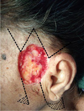
A 72-year-old female came to the dermatovenereology clinic with a brownish lump on her lateral cheek. The lump started as a mole which started to grow in the last 5 years. The lump was pruritic, bled easily and frequently produced pus. Physical examination revealed a 5 x 5 cm hyperpigmented plaque on the left lateral cheek. From anamneses and physical examination, a diagnosis of pigmented basal cell carcinoma was suspected. Laboratory examination showed that complete blood count, liver function test, kidney function test, blood glucose, electrolyte, bleeding time, clotting time were within normal limit.
Wide surgical excision of the lesion was performed followed by defect closure using advancement flap technique after all margins had been confirmed to be tumor-free. Surgical excision procedure was done with the patient in supine position. A marker was made surrounding the lesion with a margin of 0.5 cm. The operating field was disinfected using 10% povidone iodine and covered with sterile drape. One vial of Pehacaine® (lidocaine HCl 20 mg, Adrenalin 12,5 g.) was injected for local anesthesia. An incision was then performed along the marker. Bleeding was controlled and the excision wound was covered using antibiotic mesh and sterile gauze. Small samples were taken from four corners of the lesion and sent for histopathology assessment.
One week after the excision and after histopathological examination had confirmed that all borders were already tumor-free, defect closure was performed using advancement flap technique (Figure 1).
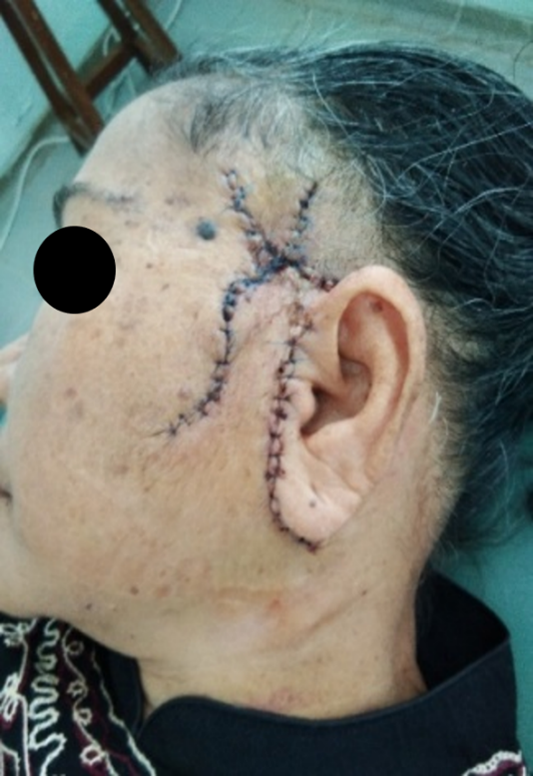
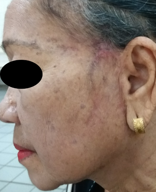
Elongated incision was done towards the frontal region and the left infra-auricular region along the pre-designed marking. This was followed by releasing the underlying tissue. Tissue that has been released were then shifted superiorly and medially towards the primary defect to close the defect. Post-operative day-6 (Figure 2) showed no sign of complications. Follow-up after three months showed excellent cosmetic and functional result (Figure 3).
DISCUSSION
Basal cell carcinoma (BCC) is the most common type (75%) of nonmelanoma skin cancer (NMSC) followed by squamous cell carcinoma (15%).(Erkul E, 2016, Bastos et al., 2015) It is a slow-growing tumor which rarely metastasizes but can cause substantial morbidity and cosmetic complaints due to its location on the face. (Trakatelli et al., 2014)
BCC is often diagnosed in patients with recurrent fragile and easy-to-bleed lesion which kept on recurring. Other characteristics include translucency, ulceration, telangiectasia, rolling border and pigmentation. Characteristics vary between different clinical subtypes, which are nodular, superficial, morphea forms, and pigmented BCC. Histopathologically, melanin pigment could be found in 30% of cases. Pigmented BCC is more frequently found in people with darker skin tone. (Firnhaber, 2012, Trakatelli et al., 2014, Chung, 2012)
When treated properly, BCC has a good prognosis. The type of skin surgical procedure differs in each case. A good surgeon has to carefully weigh the risk and benefits of each available procedures and tackle the complications that might arise. In this patient, modified Mohs micrographic procedure, which is the most effective technique for removing BCC, was done. Other than low recurrence rate, this technique provides microscopic control of the margins resulting in more accurate differentiation with surrounding normal tissues.(Firnhaber, 2012, Chung, 2012)
The defect resulting from the excision procedure was closed using combination of multiple advancement flaps. Flap procedure is done by displacement of skin tissues. This procedure is indicated if primary healing is not expected to repair the defect properly due to the size of the defect or because of high tension of the surrounding tissues. If primary closure is forced to be done, imperfect healing might result in functional or cosmetic disturbances.
The tumor was located on the lateral cheek which covered a large portion of the face and is one of the predilection sites for skin malignancy. Nearby structures such as the nose, lips and orbital region as well as the defect size need to be evaluated upon devising the reconstruction plan. If inadequate attention is given to these surrounding structures, high skin tension and excessive scar tissues formation might occur and alter the wound healing process.(Erkul E, 2016) Reconstruction in this area is particularly challenging due to its lack of laxity(Rapstine et al., 2012) and the ear, which lies just lateral to the lesion, does not provide a source for tissue flap (Bhrany et al., 2014).
There are various types of flap techniques. Compared to other flap techniques, advancement flap is the oldest and most straightforward type of adjacent tissue transfer in cutaneous surgery. This technique is done by sliding the skin flap over the surgical defect in a single vector direction while the surrounding tissues provide opposing force upon closure of the flap. This technique is also called as U-plasty or rectangular flap due to its design which extends two parallel incision from one side of the surgical defect causing one edge of the defect to become a rectangular shaped advancement flap. (Rohrer et al., 2017,Goldman G, 2012, Rao and Shende, 2016)
Advancement flap results in prominent horizontal line, making it an indispensable and more aesthetic tool in many cases of reconstruction of the eyebrow, forehead areas, upper lip and dorsal nose. One of the advantage of this technique is the ability to shift the standing cone to a more favorable location and not necessarily adjacent to the defect. (Firnhaber, 2012, Rohrer et al., 2017, Roozeboom, 2016, Goldman G, 2012) In this case, the standing cone was made posterior to the ear to hide the resulting defect. The incision lines were also made along the RSTL and preauricular crease to allow cosmetically sound result.
Although most authors advocate the use of cervicofacial flap in large lateral cheek defects (greater than 3 cm)(Bhrany et al., 2014, Cass and Terella, 2019, Rapstine et al., 2012), we opted to combine multiple advancement flaps instead of the former. Although our technique might have resulted in the formation of more vertical scars, the lateral side of the cheek is less prominent to viewers compared to other sites and hence tolerate more vertical scars.(Cass and Terella, 2019) In addition, because we utilized skin donor from area superior to the defect, the need for a long cervicofacial incision line might be avoided. Follow-up result after 3 months showed an excellent aesthetic and functional result.
This case report showed that large lateral cheek defect closure using multiple advancement flaps resulted in a cosmetically and functionally satisfying result.
Acknowledgement
The authors thanked Ivan Kurniadi, MD for his technical assistance.
Ethical Aspects
The authors declare that this work was done with the informed consent of the patient who authorized the use of the clinical photographs presented.
Funding
None to be declared
Conflict of Interest
None to be declared
Authorship Contribution
Siswanto Wahab conducted the surgical procedure, literature review, wrote the manuscript, and approved the article
Khairuddin Djawad conducted the surgical procedure, reviewed the manuscript, and approved the article
REFERENCES
Bastos, TC., Uribe, NC., Brandão, CM., de Carvalho, MM. 2015. Unipedicled advancement flap from the lower cheek for the reconstruction of a large nasal surgical defect after the excision of a basal cell carcinoma. Surg Cosmet Dermatol, 7(4): 346-349, http://dx.doi.org/10.5935/scd1984-8773.201574626.
Bhrany, AD., Bradley, DT., Murakami, CS. 2014. Reconstruction of the cheek. In: Baker, S. R. (ed.) Local Flaps in Facial Reconstruction. 3rd ed. Philadelphia: Elsevier Saunders.
Cass, ND., Terella, AM. 2019. Reconstruction of the Cheek. Facial Plastic Surgery Clinics, 27(1): 55-66.
Chung, S. 2012. Basal Cell Carcinoma. Archieve of plastic surgery, 39(2): 166-170.
Erkul, PK., Day, T. 2016. Surgical Planning for Resection and Reconstruction of Facial Cutaneous Malignancies: Original article. Int. J. Head Neck Surg., 7: 149-164. https://www.ijhns.com/doi/IJHNS/pdf/10.5005/jp-journals-10001-1281 .
Firnhaber, JM. Diagnosis and treatment of basal cell and squamous cell carcinoma. 2012. Am. Fam. Physician, 86(2): 161-168. https://www.aafp.org/afp/2012/0715/p161.html
Goldman, DL., Yelverton, C. 2012. Facial Flaps Surgery, McGraw-Hill Education.
Quinn, AG., 2010. Rook's Text Book of Dermatology., West Sussex-UK, Wiley-Blackwell.
Rao, JK., Shende, KS. 2016. Overview of local flaps of the face for reconstruction of cutaneous malignancies: single institutional experience of seventy cases. J. Cutan. Aesthet. Surg., 9(4): 220 https://doi.org/10.4103/0974-2077.197029 .
Rapstine, ED., Knaus, WJ., Thornton, JF. Simplifying cheek reconstruction: a review of over 400 cases.2012. Plast. Reconstr. Surg., 129(6): 1291-1299 https://doi.org/10.1097/prs.0b013e31824ecac7.
Rezakovic, S, Zuzul, K., Kostovic, K. Basal cell carcinoma: review of treatment modalities. 2014. J Dermatolog Clin Res, 2(5): 1035. https://www.jscimedcentral.com/Dermatology/dermatology-2-1035.pdf .
Rohrer, TE., Cook, JL., Kaufman, A. 2017. Flaps and Grafts in Dermatologic Surgery E-Book, Elsevier Health Sciences.
Roozeboom, M. 2016. Diagnosis and treatment of Basal Cell Carcinoma, Maastricht, Universitaire Pers Maastricht.
Tang, J. Y., Epstein Jr, E.H.; Oro, A.E. 2019. Basal Cell Carcinoma and Basal Cell Nevus Syndrome. In: Kang, S., Amagai, M., Bruckner, A. L., Enk, A. H., Morgolis, D. J., McMichael, A. J. & Orringer, J. S. (eds.) Fitzpatrick's Dermatology 9th Ed. McGraw-Hill Education.
Trakatelli, M., Morton, C., Nagore, E., Ulrich, C., Del Marmol, V., Peris, K., Basset-Seguin, N. 2014. Update of the European guidelines for basal cell carcinoma management. Eur. J. Dermatol.,
Author notes
duddin@ymail.com
Additional information
How to cite: Wahab, S., & Djawad, K. 2021. Large Basal Cell Carcinoma Defect Closure on The Lateral Cheek Using Multiple Advancement Flaps. International Journal of Medical and Surgical Sciences, 8(1), 1-7. https://doi.org/10.32457/ijmss.v8i1.671
Alternative link
https://revistas.uautonoma.cl/index.php/ijmss (html)
https://revistas.uautonoma.cl/index.php/ijmss/article/view/671 (html)
https://revistas.uautonoma.cl/index.php/ijmss/article/view/671/1074 (pdf)